

TECHNOLOGY - GraFuture™
B A C KReduced Graphene
Oxide Grid
To overcome the bottleneck in cryo-EM sample preparation, we developed a new type of reduced graphene oxide grid (GraFuture™), using it to achieve a better reproducibility and efficiency of specimen preparation.

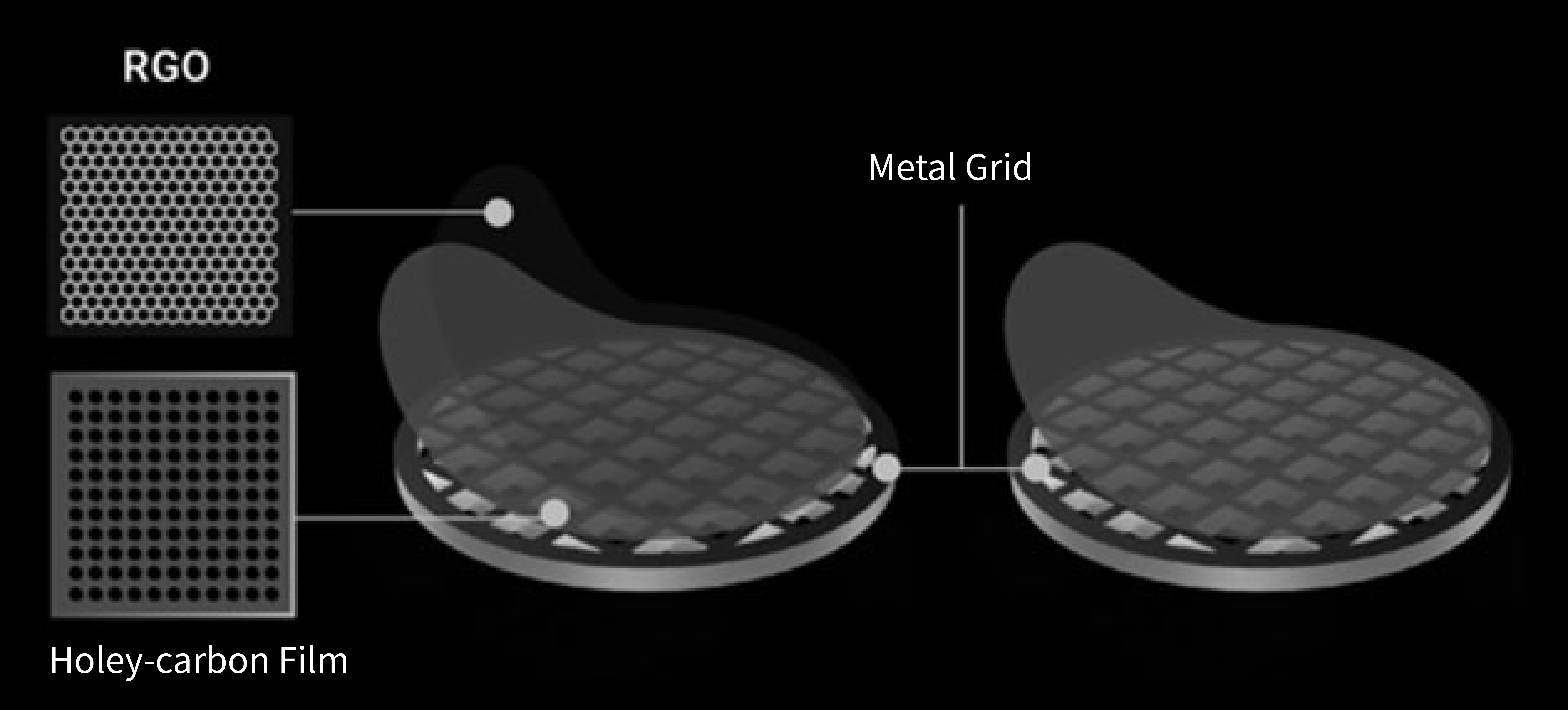
Fig1. Schematic diagram of EM grids coated with reduced
graphene oxide¹ (left) and conventional ones² (right).
A big leap can be taken in structure determination once good cryo-EM grids are prepared.
The behavior of molecular embedded in amorphous ice plays an important role in the best achievable resolution for cryo-EM structure determination, leaving specimen preparation to be a major bottleneck limiting in this method .
Aiming to improve reproducibility and controllability, we developed a facile and robust strategy to use reduced graphene oxide(RGO) membrane as supporting film in cryo-EM specimen preparation. This grid has been used in our platform and showed great power to reconstruct biomolecules (including sub-100-kDa molecules), even at near-atomic resolution that can not be reached by conventional used grid.

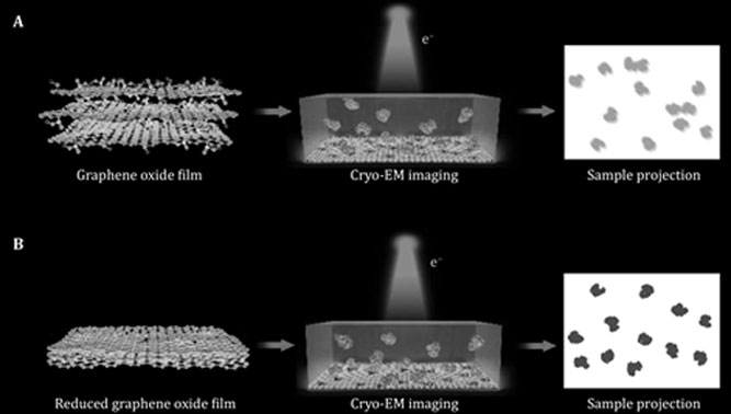
Fig2. Difference in Graphene oxide (A) and reduced
graphene oxide (B) for single-particle cryo-EM analysis.
GraFuture™: offering promise for challenging samples that are
inaccessible by cryo-EM before.
Compared with conventional supporting films, EM grids coated with our RGO membrane was characterized with several superior properties.
- Decreased interlayer space results in thinner ice which is just thick enough to support the particles. Lower background noise significantly improves the image quality.
- Improved biocompatibility and nice particle-absorption ability not only enhances particle concentration, but also alleviates air water interface problem which is hypothesized to cause denaturation and preferred orientation.
- Enhanced electrical/thermal conductivity contributes to no visible beam-induced footprints and minimized radiation damage.
- High mechanical strength means adequate strength to support the ice layer and reduced beam-induced motion.
Using our grids, boundaries are continuously being pushed in structure determination.
- Small molecules (sub-100 kDa)
- Low protein concentration (~100 nM)
- Fragile and sensitive proteins
- Preferential orientation on commercially purchased grids

Fig2. Difference in Graphene oxide (A) and reduced
graphene oxide (B) for single-particle cryo-EM analysis.
Featured Publication
Mechanism of spliceosome remodeling by the ATPase/ helicase Prp2 and its coactivator Spp2
Rui Bai et al. SPA & RGO grid DOI: 10.1126/science.abe8863
All major states of assembled spliceosome have been determined at near-atomic resolution, however, none of the ATPase/helicases in the presence of the spliceosome has been visualized in atomic details. Prof, Yigong Shi's laboratory has successfully resolved the cryo-EM structures of the S. cerevisiae Bact complex which is absolutely an amazing work. By comparing the structures of Prp2 before and after recruitment into the activated spliceosome, it has been addressed how spliceosome remodeling occurs. Specifically, RGO grid is used for cryo-EM specimen preparation during EM data acquisition for Prp2 (molecular weight is only 100 kDa).
V i e wReduced graphene oxide membrane as supporting film for high-resolution cryo-EM
Nan Liu et al. SPA & RGO grid DOI: 10.52601/bpr.2021.210007
Although single-particle cryo-EM is rapidly becoming an attractive technology in the field of structure biology, reliable and versatile specimen preparation can be time consuming and labor intensive. To achieve more reproducible and desirable particle behavior, Hongwei Wang's laboratory has developed a robust method to generated EM grids coated with reduced graphene oxide membrane in large batch for high-resolution cryo-EM structural determination. RGO supporting film is particularly useful in reconstruction of sub-100-kDa biomolecules at near-atomic resolution, as exemplified by the reconstruction results of RBD-ACE2 complex.
V i e w